
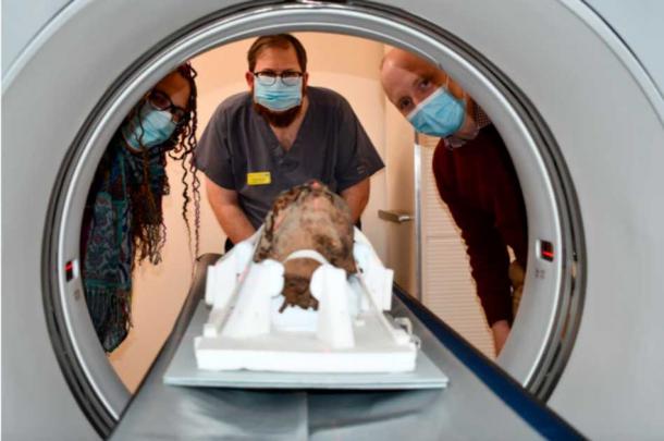
Archaeologists and hospital staff in England have teamed up to scan an ancient Egyptian mummy’s head found in an attic. The head, dating back 2,700 years, was shipped as a souvenir in the 1820s to Ramsgate, a seaside town in East Kent. After the homeowner’s death, the head, encased in a glass display, was donated by his brother to the Canterbury Museums and Galleries collection.
The Discovery Process
Craig Bowen, the galleries collections and learning manager at Canterbury Museum, explained that the head was found by a man who inherited it from his brother, who had received it from a “Dr. Coates” in the early to mid-twentieth century. Initial X-rays conducted in 2020 at Canterbury Christ Church University revealed the mummy was an adult female. However, a recent detailed CT scan has provided more data about the individual before her death. CT scans involve taking multiple X-ray images from different angles, which are then combined to create a high-resolution map of the body.
Brainless in the Afterlife
James Elliott, a lecturer in diagnostic radiography at Canterbury Christ Church University and senior radiographer at Maidstone and Tunbridge Wells NHS Trust, conducted the new CT scans. These scans offered a wealth of information, including details on dental status, pathologies, and methods of preservation, helping estimate the age and sex of the mummy. Notably, the scans revealed that the woman’s brain had been removed, indicating she underwent the traditional purification and mummification procedures in a Per-Nefer, or ‘house of beauty.’
Insights into Ancient Egyptian Practices
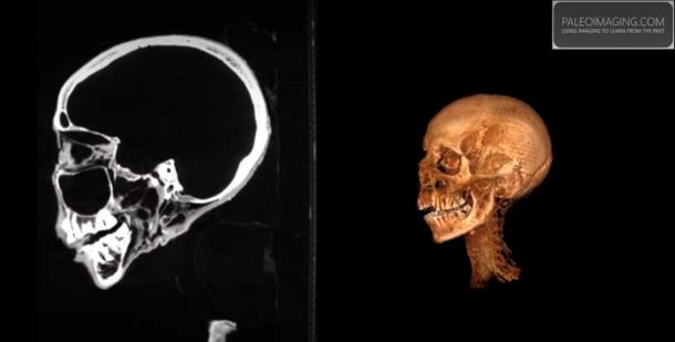
The embalmers typically used tools like hammers, chisels, and iron hooks to remove the brain through the nasal bone, followed by washing the cranial cavity with water. Craig Bowen noted the irony in the ancient Egyptians’ belief that a person’s mind resided in the heart, not the brain, which they regarded as insignificant.
Further Discoveries and Future Plans
The CT scans also revealed that the woman’s tongue was well-preserved and her teeth were worn, suggesting a diet of coarse foods. Interestingly, a tubing of unknown material was found in her left nostril and spinal canal. The origins and composition of these materials remain a mystery and could potentially be modern additions, perhaps from the Victorian era.
Bowen stated that this project aims to preserve the head and display it in conservation-grade packaging for public viewing. The team plans to use the CT scanning data to create a three-dimensional replica of the woman’s head. They will also attempt to reconstruct her face in 3D, allowing for a visual representation without exposing the actual artifact. This effort will help bring to life the history and culture encapsulated in this ancient Egyptian relic.